An intraventricular fetus-in-fetu, a malformed monochorionic diamniotic twin, was identified in a 1-year-old girl with motor delay and enlarged head circumference (Figure 1). After surgical removal, whole-genome sequencing revealed identical single-nucleotide variants in the host child and fetus-in-fetu, with extensive de novo copy number gains in the fetus-in-fetu (Figure 2, eMethods, links.lww.com/WNL/C529), suggesting the significance of copy number variation during embryogenesis.
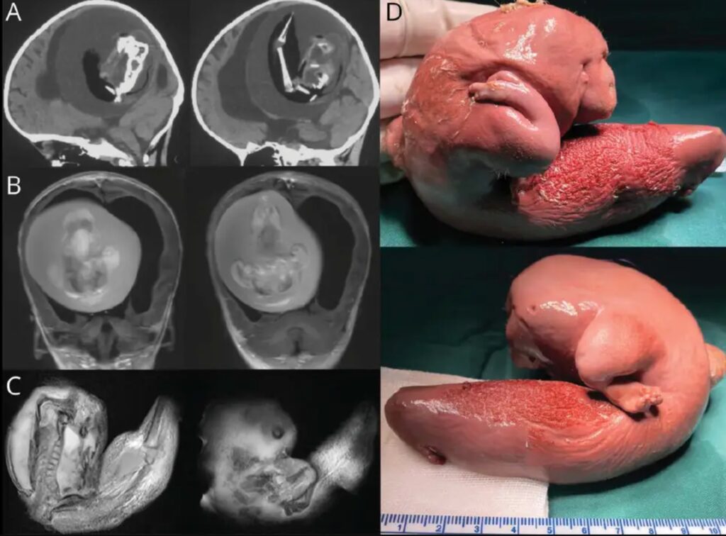
Figure 2 Copy Number Duplication in Fetus-in-Fetu

The intracranial fetus-in-fetu is proposed to arise from unseparated blastocysts. The conjoined parts develop into the forebrain of host fetus and envelop the other embryo during neural plate folding.1 Fetus-in-fetu can be distinguished from teratomas based on the younger age of presenting patients and the presence of vertebrae or internal organs.2
Footnote
Teaching Slides links.lww.com/WNL/C530
References
1.Miura S, Miura K, Yamamoto T, et al. Origin and mechanisms of formation of fetus-in-fetu: two cases with genotype and methylation analyses. Am J Med Genet A. 2006;140A(16), 1737-1743.
2.Huddle LN, Fuller C, Powell T, et al. Intraventricular twin fetuses in fetu. J Neurosurg Pediatr. 2012;9(1), 17-23.